


When the Body Changes: Body Shape Analysis Reveals What Really Changed
Our bodies are constantly changing.
Sometimes we notice it in the mirror. Sometimes in the way clothes fit. Sometimes in a photograph taken months apart.
A smoother thigh. A different waistline. A firmer contour. A subtle change in shape after a cosmetic product or aesthetic treatment.
But seeing a difference and scientifically demonstrating a difference are not the same thing.
Was the circumference actually reduced?
Did the volume change?
Did the body contour really reshape?
Are cellulite dimples less pronounced?
And perhaps most importantly: did the improvement last?
As cosmetic and aesthetic treatments become increasingly sophisticated, so does the need to evaluate their results.
Because when the body changes, the real scientific question isn't simply whether we can see it.
It's whether we can measure it.
The Body Changes. For Many Different Reasons.
Body shape is not static.
It evolves naturally throughout life under the influence of age, weight fluctuations, hormonal changes, pregnancy, lifestyle, physical activity and many other factors.
Some changes affect overall body morphology. Others are highly localized.
The abdomen may change in volume.
The waistline may change in circumference.
The contours of the thighs or buttocks may evolve.
Skin may appear firmer or looser.
And its surface may develop the characteristic depressions, elevations and irregularities associated with cellulite.
These changes are different, but they have something important in common:
They modify the body's three-dimensional morphology.
And many of them have become important targets for the cosmetic, dermocosmetic and aesthetic industries.
From Cellulite to Body Contouring: What Are We Trying to Change?
Body care has moved far beyond simple moisturization.
Today's cosmetic formulations, aesthetic technologies and treatment protocols may target very specific aspects of body appearance.
Cellulite
Cellulite is characterized by an uneven, dimpled skin surface, most commonly affecting areas such as the thighs and buttocks. Its appearance is associated with complex interactions between the skin, connective structures and underlying adipose tissue.
What we ultimately see at the surface are topographical irregularities: depressions, elevations, dimples and nodules.
And those surface characteristics can be studied quantitatively.
Research has demonstrated that three-dimensional skin surface topography can be used to characterize cellulite morphology, with specific surface parameters correlating with expert assessments of cellulite severity.
So rather than simply asking: "Does the cellulite look better?" researchers can investigate: "How did the surface morphology change?"

Slimming
A slimming effect presents a different measurement challenge.
Here, researchers may be interested in changes in the dimensions of a specific body area.
Has the circumference of the thigh changed?
Has abdominal volume decreased?
How large is the difference?
And is that difference consistent across repeated measurements?
A measuring tape may provide a circumference. But the body isn't a perfect cylinder, and slimming doesn't necessarily occur uniformly around an anatomical region.
Understanding the result may therefore require looking beyond a single number.

Body Contouring
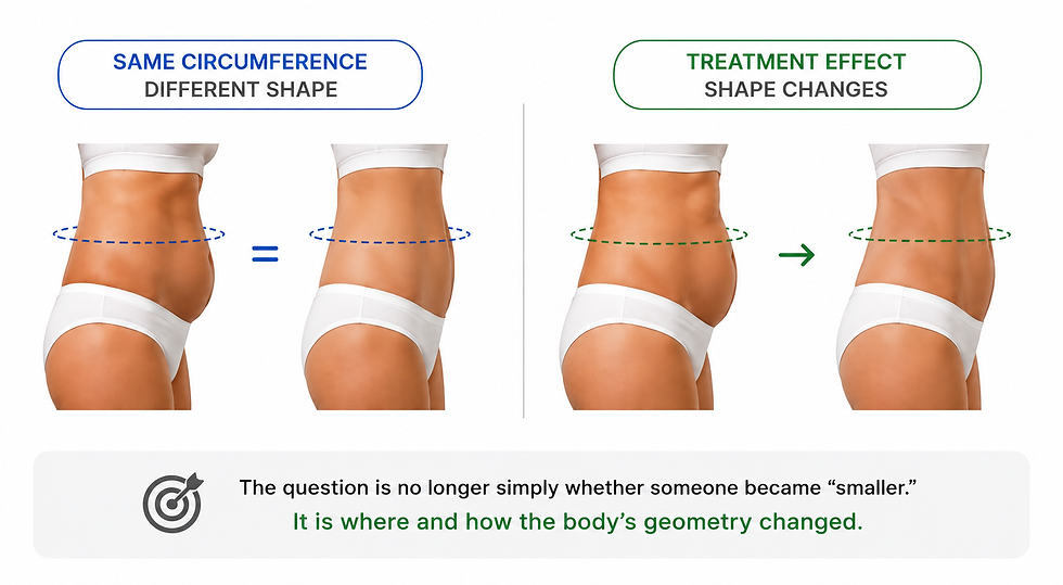
Body contouring adds another dimension: shape.
Two body areas could potentially have similar circumferences while presenting different contours.
A localized treatment may alter the morphology of the abdomen, thigh, hip or another area without producing a dramatic change in overall body weight.
For body-contouring research, the relevant endpoints may therefore include: Shape. Volume. Circumference. Localized morphological change.
The question is no longer simply whether someone became "smaller."
It is where and how the body's geometry changed.
Firming & Reshaping
Firming and reshaping treatments can produce subtler visual changes.
The objective may not necessarily be substantial volume reduction.
Instead, researchers may want to determine whether a treated area presents a measurable morphological difference compared with baseline.
Has the contour changed?
Has the shape evolved?
Can the effect be localized?
And can that change be detected consistently over time?
Different treatments therefore require different endpoints. But they all lead to the same challenge:
How do we turn a visible change into measurable evidence?

What We See Isn't Always What We Can Measure
Before-and-after photography remains extremely valuable. It documents appearance. It communicates results intuitively. And it allows researchers, clinicians and consumers to visually compare two moments in time.
But a photograph is still a two-dimensional representation of a three-dimensional body.
Lighting can change. Posture can change. Camera position can change. Body orientation can change.
And even subtle differences between acquisitions may influence how contours appear.
Traditional circumference measurements provide quantitative information, but they introduce another challenge: repositioning the measurement at exactly the same anatomical location at every visit.
Scientific research comparing manual circumference measurement with 3D surface imaging has found greater reproducibility with 3D measurement for thigh and abdominal circumference.
This doesn't make photographs or measuring tapes irrelevant. Far from it.
It means that when the expected changes are small, localized or progressive, additional objective measurement can strengthen the evaluation.
The Body Is 3D. Shouldn't Its Changes Be Measured in 3D?
Consider the shape of a thigh. Or an abdomen. Or a hip. Its geometry exists simultaneously in height, width and depth.
Cellulite adds another level of complexity because the feature of interest is the surface relief itself.
Flatten that information into a conventional photograph and some of its three-dimensional characteristics inevitably become harder to quantify.
This is why body shape analysis is increasingly interesting for cosmetic and aesthetic research.
Instead of reducing the body to a visual impression or isolated manual measurement, researchers can capture its surface geometry and convert physical changes into quantitative data.
The question changes from: "Can we see a difference?" to: "Can we quantify the difference?"
One Treatment. Several Visits. Which Result Tells the Real Story?
There's another challenge. Treatments happen over time. So do their results.
Imagine a body-contouring or anti-cellulite study:
Baseline → Week 4 → Week 8 → Week 12 → Follow-Up
A participant may show little visible change initially. The effect may then progressively increase. It may reach a plateau. Or the improvement observed at the end of treatment may partially diminish after treatment stops.
If researchers measure only the beginning and the end, they see two snapshots. If they perform longitudinal body shape analysis, they can investigate the evolution of the response.
This is particularly important when the scientific question is not merely: Did the treatment work?
but also:
When did the change appear?
How large did it become?
Did it continue to improve?
Was it maintained?
Published cellulite research illustrates the value of this approach. Three-dimensional imaging has been used at multiple time points to quantify changes in cellulite dimples and evaluate whether improvements persisted months after treatment.
A treatment result isn't just a before and an after. It's an evolution.
Why Accurate Body Shape Analysis Matters to the Cosmetic Industry
For a consumer, an improvement may simply mean:
"My skin looks smoother."
"My waist looks more defined."
"My thighs look different."
These perceptions matter.
But cosmetic companies, research laboratories and CROs need to go further. They need to determine whether the observed effect can be objectively demonstrated. That makes accurate body measurement relevant throughout the development process.
Research
Objective measurements help researchers understand how body morphology and surface topography respond to products and treatments.
Product & Treatment Development
Quantitative endpoints can help compare formulations, protocols, application conditions or treatment approaches.
Efficacy Testing
Measurements of volume, circumference, shape or surface characteristics can complement photography, clinical grading and participant questionnaires.
Clinical Studies
Standardized measurements make it possible to compare the same anatomical region across study visits and quantify changes from baseline.
Claim Substantiation
When supported by an appropriately designed study, quantitative measurements can contribute objective evidence for claims associated with measurable effects such as changes in circumference, body contours or cellulite appearance.
Scientific & Marketing Communication
Numbers provide evidence. Images provide understanding.
Combining quantitative results with intuitive three-dimensional visualizations can make study outcomes easier to interpret and communicate across R&D, clinical, regulatory and marketing teams.
The objective isn't to transform science into marketing. It is to give marketing stronger science to communicate.
Cellulite Is a Perfect Example: Don't Just Grade It. Measure Its Surface.
Cellulite demonstrates particularly well why three-dimensional surface measurement matters.
The characteristic appearance of cellulite is fundamentally related to surface topography.
Dimples form depressions. Nodules and elevations contribute to irregular surface relief. The resulting "orange-peel" appearance isn't simply a color difference, it has geometry. Traditional clinical grading can classify severity.
Photography can document appearance. Subject questionnaires can capture perceived improvement.
But 3D surface analysis adds another layer: quantitative surface statistics.
Research has demonstrated the scientific value of this approach. Three-dimensional methods have quantified changes in cellulite dimple depth and negative volume after aesthetic treatments, providing objective numerical endpoints alongside visual and clinical assessments.
And 3D technology itself has been used in published research to capture cellulite-related 3D surface structures and quantify changes in dimple volume over time.
This allows cellulite efficacy assessment to move from:
"The surface looks smoother."
toward:
"The surface morphology measurably changed."
From Visual Assessment to True 3D Body Measurement
This is where True 3D imaging becomes particularly relevant.
A True 3D measurement system captures the actual surface geometry of the body and reconstructs it as quantitative three-dimensional data.
Instead of relying exclusively on a photograph, researchers obtain a measurable 3D representation of the anatomical area.
For the applications we've discussed, that can provide several highly relevant endpoints:

Suddenly, a body change isn't simply visible. It has dimensions.
Introducing AEVA-HE²:When Body Changes Become Quantitative |
EOTECH's AEVA-HE² brings high-resolution True 3D imaging to the objective assessment of body morphology and skin surface relief.
Based on fringe projection combined with stereometry, AEVA-HE² captures the three-dimensional geometry of the measured surface. Different fields of view allow the technology to be adapted to different measurement areas, including body applications.
EOTECH specifically identifies volume, circumference, cellulite and microrelief among its body applications.

Combined with the AEVA software, the acquired 3D data can be analyzed quantitatively rather than evaluated solely as images.
This means researchers can investigate endpoints directly related to the objective of the study.
For Cellulite
The three-dimensional skin surface can be analyzed through surface statistics associated with dimples and nodules, providing quantitative information about the morphology of the cellulite-affected area.
For Slimming
Measurements of circumference and volume provide quantitative endpoints for evaluating dimensional changes in a selected body area.
For Body Contouring
Shape, volume and circumference can be evaluated to characterize how the morphology of a treated area changes.
For Firming & Reshaping
3D comparison can reveal morphological changes that may be difficult to describe through a conventional photograph or isolated measurement alone.
And importantly, these measurements don't have to stop at a single before-and-after comparison.
Same Body Area. Same Position. Different Time.
For longitudinal studies, measurement accuracy isn't enough.
Reproducibility matters.
If a volunteer stands differently between two acquisitions, apparent changes in body shape may reflect positioning rather than treatment.

That is why standardized repositioning becomes critical.
EOTECH's VisioHOP positioning system is designed for reproducible face and body measurements with AEVA-HE². In body mode, the volunteer is measured standing while the system automates sensor positioning around the subject, enabling measurements from different positions and supporting 360° acquisition.
This makes it possible to return to the same measurement conditions across study visits and compare the evolution of body morphology more consistently.
Baseline → Treatment → Follow-Up → 3D Comparison
The objective is not merely to create a visually impressive 3D body image. It is to create comparable quantitative data over time.
When the Body Changes, Measure What Really Changed.
The human eye is remarkably good at noticing differences.
But scientific research needs to go further.
When evaluating cellulite, slimming, body contouring or reshaping treatments, the important question isn't only whether a participant appears different. It's: What changed?, where?, by how much?, and did that change persist over time?
True 3D body measurement gives researchers a way to answer those questions quantitatively.
From cellulite surface statistics to body circumference, volume and morphology, it transforms visible changes into measurable scientific endpoints.
Because the body isn't two-dimensional. And neither are its changes.

AEVA-HE²
Captures body surface geometry in high-resolution 3D using fringe projection combined with stereometry.
Quantifies volume and changes in volume for selected body areas.
Calculates circumference directly from the captured 3D geometry for objective dimensional assessment.
Quantifies 3D shape changes to evaluate morphological differences in treated body areas.
Provides surface statistics for cellulite dimples and nodules to quantitatively characterize skin-surface irregularities.
Allows defined regions of interest to be extracted and analyzed independently for targeted assessment.
Compares 3D acquisitions to quantify shape and volume differences between measurement time points.
Manages measurements across multiple study time points, enabling quantitative evaluation of how body morphology evolves throughout a study.




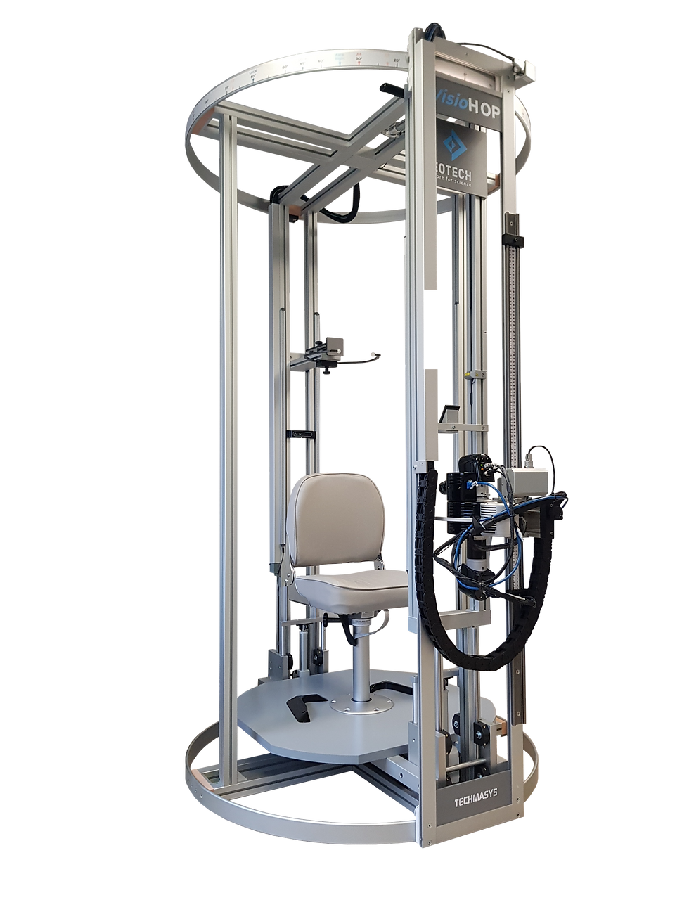
VisioHOP
REPRODUCIBLE POSITIONING
Consistent volunteer positioning across study time points.
360° BODY ACQUISITION
Automated measurement around the body from multiple orientations.
AUTOMATED SENSOR MOVEMENT
Controlled vertical and angular positioning of AEVA-HE².
SUBJECT STABILIZATION
Helps maintain a stable body position throughout the acquisition sequence.
LONGITUDINAL CONSISTENCY
Supports comparable baseline and follow-up measurements for quantitative 3D analysis.
Works with AEVA software and AEVA-HE² as an integrated acquisition workflow, coordinating sensor movement and the positions required for the study.
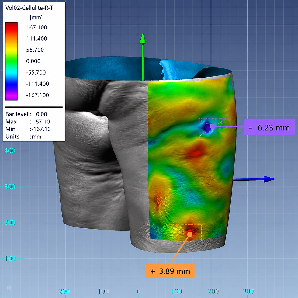
AEVA Software
AUTOMATED 3D ANALYSIS
Processes acquired 3D data into quantitative body measurements and visual results.
BODY VOLUME & CIRCUMFERENCE
Quantifies volume and circumference from captured body geometry.
SHAPE CHANGE ANALYSIS
Compares 3D surfaces to quantify morphological changes between acquisitions.
CELLULITE ANALYSIS
Provides surface statistics for cellulite dimples and nodules, including localized surface analysis.
REGION OF INTEREST (ROI)
Selects and analyzes specific body areas independently for targeted evaluation.
3D COMPARISON MAPS
Visualizes differences between 3D acquisitions using statistical deviation and pseudo-color displays.
LONGITUDINAL STUDY MANAGEMENT
Organizes measurements across volunteers and study time points for treatment follow-up.
QUANTITATIVE + VISUAL RESULTS
Generates calculated values, charts, 3D models and visualizations for interpretation and reporting.
You See the Body Change.
3D Body Imaging Shows You What Really Changed.
Discover EOTECH’s full range of skin research Instruments at Skinlabs and see how advanced measurement tools help you do more for science. |
PUBLICATIONS |


Comments