


3D Surface Microstructure Measurement for Advanced Biological Research
- Emma Danciu
- Feb 1
- 7 min read
Updated: Feb 3
The skin and nails are complex biological structures whose function and appearance are defined by microscopic architecture. Changes in microstructure influence barrier performance, hydration, mechanical strength, penetration, aging, and visible surface quality.
Accurate measurement of skin and nail microstructure—whether performed in-vivo, ex vivo, or in-vitro—is fundamental for cosmetic science, pharmaceutical development, biomedical engineering, and dermatological research. These measurements transform subtle biological changes into objective, quantifiable data that drive innovation, product validation, and regulatory compliance.
Why Accurate Surface Microstructure Measurement Matters
— for Science and Industry
Surface features such as microrelief, fine lines, pore topography, nail microstructure, and changes induced by treatments are crucial biomarkers of health, aging, and treatment efficacy.
To fully understand these surface microstructure biomarkers, it is essential to measure them across multiple biological models.
In vivo, ex vivo, and in vitro surface microstructure measurements each provide distinct and complementary perspectives on skin and nail structure, function, and response to intervention. Together, these measurement models enable comprehensive evaluation of surface morphology, micro texture, and microrelief, supporting robust research, product development, and clinical validation.
Below is a detailed presentation of each of these experimental research models.
In‑Vivo Measurements
They capture real biological variability under natural physiological conditions — essential for evaluating nail health and integrity (e.g., nail thickness, keratin structure changes), monitoring skin elasticity, hydration, and microrelief with clinical relevance, and supporting longitudinal studies tracking treatment effects.
In-vivo skin and nails measurement technologies are essential for clinical trials, providing accurate, real-time data directly from living tissue. They enable continuous monitoring of nail and skin health over time, detecting subtle changes that support personalized care and treatment optimization.
Additionally, in-vivo measurements serve as non-invasive biomarkers for systemic diseases, offering critical insights into overall health without invasive procedures.

Ideal for dermatology (monitoring nail health), cosmetic research (developing fortified nail treatments), and pharmaceutical research (for drug safety and side-effect profiling).
Ex‑Vivo Measurements
precise quantification of collagen/elastin organization and tissue architecture, measures epidermal thickness, detects skin aging and damage mechanisms, and evaluation of fine surface texture changes not visible to the naked eye.
Ex-vivo skin and tissue measurements are powerful tools for anti-aging and regenerative product validation, providing precise data on efficacy before clinical use. They are essential for studying disease mechanisms, including eczema, scarring, and psoriasis, allowing researchers to investigate cellular and tissue-level changes in a controlled environment.
Ex-vivo testing also supports toxicology assessments, ensuring the safety of new compounds and formulations. These approaches deliver accurate, reproducible, and high-resolution insights.

Invaluable for academic and clinical research (studying skin physiology and pathology), cosmetic science and skincare innovation (developing creams, serums, and anti-aging products), and academic and clinical research centers (studying skin physiology and pathology).
In-Vitro Measurements
In-vitro studies use isolated tissues or reconstructed skin models, offering controlled, reproducible measurements.
Controlled lab models let researchers screen products before human trials, quantify pores, fine lines, surface roughness consistently across samples, and cellular responses, eliminate ethical and physiological constraints of live testing.
Ex-vivo skin testing is a critical step for screening anti-aging compounds before clinical trials, ensuring only the most promising formulations advance. It allows researchers to assess irritation or allergenic potential, improving safety and reducing risks in human studies.
Moreover, ex-vivo measurements help in optimizing hydration, elasticity, and skin texture formulations, providing precise, reproducible data to guide product development.

Optimal for cosmetic formulators (innovating new serums, masks, and moisturizers), pharmaceutical research teams (for dermatological drug development), and regenerative medicine (focusing on tissue engineering and wound healing studies).
Why Measuring Across In-Vivo, Ex-Vivo, and In-Vitro is Essential
Measuring across in-vivo, ex-vivo, and in-vitro models is essential for a comprehensive understanding of skin and nail health. Each approach provides unique insights.
Integrating these methods supports product development and validation, informing formulation optimization, safety assessments, and efficacy verification for cosmetics, pharmaceuticals, and clinical research.
High-quality microstructure data also ensures regulatory compliance, backing scientific claims with robust evidence.
Furthermore, this multi-level approach enhances precision research, enabling longitudinal studies, reproducibility, and benchmarking critical for peer-reviewed publications and clinical studies.
Driving Innovation Across Multiple Industries
From cosmetic brands seeking to reduce fine lines to pharmaceutical companies developing topical therapeutics, and dermatology clinics monitoring skin health, precise measurement of in-vivo, ex-vivo, and in-vitro samples is indispensable.
By analyzing nails, skin microstructure, microrelief, pores, and fine lines, companies and researchers can innovate more effectively, create scientifically validated products, and ultimately enhance human health and beauty.
Reliable measurement of in‑vivo, ex‑vivo, and in‑vitro microstructures teaches us how biological surfaces behave, respond to treatments, and change over time.
To advance these fields, cutting-edge tools like EvaSURF from EOTECH deliver unmatched precision in 3D surface microstructure measurement and advanced analytical power. |


Among the 3D imaging tools available today, EvaSURF stands out as an essential instrument for in‑vivo, ex‑vivo, and in‑vitro surface measurement. It’s part of EOTECH’s advanced high‑resolution imaging family — technologies that have been scientifically validated and widely adopted in research settings worldwide.
What Is EvaSURF? |
EvaSURF is a high‑resolution 3D surface scanner engineered to capture micro‑topographies of biological surfaces with exceptional accuracy. It is specifically designed to measure microrelief, microstructures of skin and nails, pores, and fine lines.
Unlike standard 2D imaging, EvaSURF creates full 3D surface maps, enabling researchers to quantify surface shape, pattern density, roughness, and morphological changes with a level of detail that traditional photography and 2D microscopy cannot match.


The Technology behind EvaSURF |
EvaSURF is a 3D surface microstructure measurement system that combines fringe projection with stereovision.
It allows to capture highly accurate, non-contact, three-dimensional representations of skin, nails, and other biological surfaces.
By integrating these two proven optical techniques, EvaSURF achieves high spatial resolution, excellent depth accuracy, and fast image acquisition—making it well suited for in-vivo, ex-vivo, and in-vitro surface measurement in research and clinical environments.
Fringe projection works by projecting a sequence of structured light patterns onto the surface being measured. When these fringes interact with a three-dimensional surface, they become distorted according to the object’s shape. A camera records the deformed patterns, and advanced algorithms analyze phase shifts and distortions to calculate precise height values for every point on the surface.

This technique provides dense, high-resolution surface topography, capturing fine microrelief such as wrinkles, pores, ridges, and surface texture with micron-level detail.
Stereovision involves two cameras positioned at known angles, observing the same surface simultaneously—similar to how human eyes perceive depth. Each camera captures a slightly different view of the surface.
By identifying corresponding points in both images and calculating their positional differences (disparity), the system computes accurate three-dimensional coordinates.
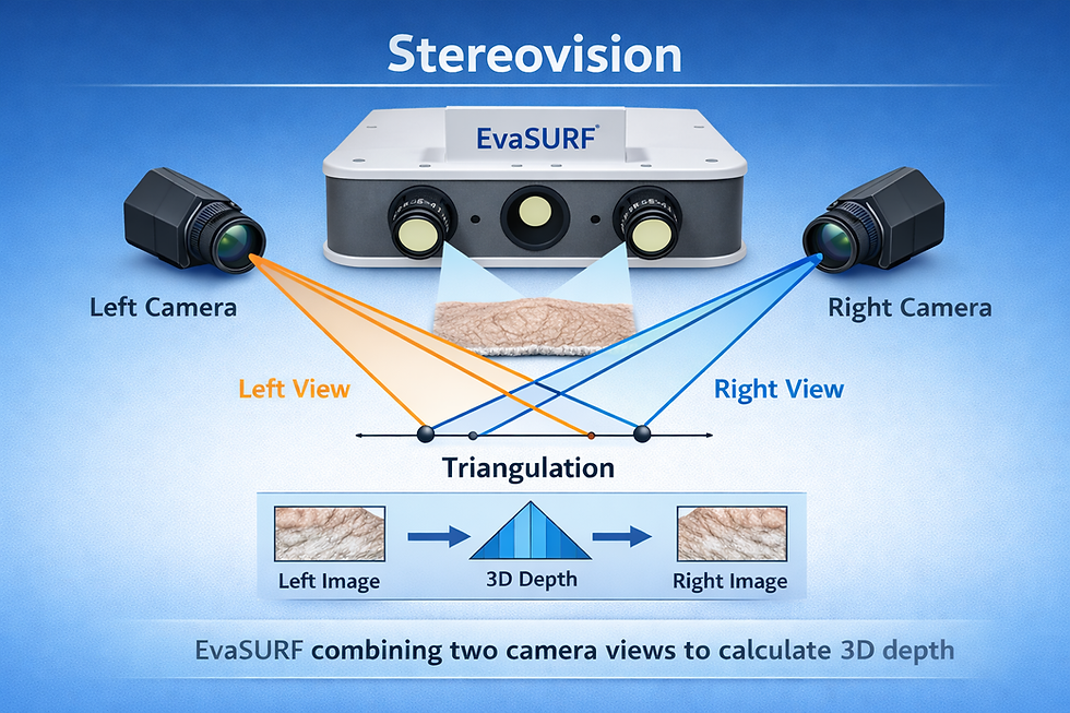
Stereovision adds robust depth perception and geometric accuracy, especially for complex or curved biological surfaces.
By combining fringe projection and stereovision, EvaSURF leverages the strengths of both techniques. Fringe projection delivers highly detailed surface texture and local height variations, while stereovision ensures precise global shape reconstruction and spatial consistency.
Together, they produce a fast, non-invasive, and highly accurate 3D surface map that supports quantitative analysis of roughness, volume, depth, and morphological changes over time. This hybrid approach makes EvaSURF a powerful tool for skin research, cosmetic efficacy studies, clinical assessments, and material surface analysis. |
Why EvaSURF Is Essential for Research |
First of all, EvaSURF delivers true 3D surface topography, using optical metrology methods that produce dense, precise 3D height maps of surfaces. This means it can resolve subtle variations in surface texture, from tiny nail irregularities to subtle skin relief patterns.
This 3D data is far more informative than flat images, allowing researchers to extract meaningful quantitative parameters, such as:
Surface, profile roughness and fine lines statistics
Feature densities (e.g., pore counts)
Exact microrelief geometry
Surface feature measurement (number, volume, area, depth, circumference)
These are indispensable in validating product claims or understanding biological aging processes.
Second, EvaSURF offers high sensitivity, detecting even the subtlest changes in skin and nail surfaces. It can detect minute changes induced by aging, treatments, or environmental exposure long before they become visually apparent.
This level of sensitivity is critical for:
Anti‑aging product efficacy studies.
Evaluating hydration or barrier changes in skin.
Tracking nail surface degradation in disease or treatment.
Such sensitivity strengthens the statistical reliability of research observations and supports more compelling scientific claims.
Third, EvaSURF demonstrates exceptional versatility across sample types, accurately scanning in-vivo live skin, ex-vivo explants or tissue samples, or in-vitro engineered replicas.
EvaSURF adapts comfortably to different workflows. This flexibility makes it ideal for:
Cosmetic efficacy testing
Biomedical research
Material science surface studies
Because it supports both clinical and lab environments, EvaSURF bridges the gap between controlled experiments and real‑world biological surfaces.
Finally, EvaSURF is fully compatible with AEVA, the advanced analysis software. The analytical engine behind EvaSURF and other EOTECH scanners — delivers powerful processing and visualization. AEVA transforms raw 3D data into interpretable results like surface roughness, pattern detection, and volumetric analysis, making it a true end‑to‑end research solution. Read more about AEVA Software.
What EvaSURF Brings to Scientific Discovery |
EvaSURF has enhanced surface measurement through several key features:
It uses scientifically validated metrology with fringe projection, ensures high reproducibility and low variability between operators for longitudinal studies and comparative trials, and provides standardized workflows to maintain consistent results from scan to scan.
Beyond visual 3D images, EvaSURF delivers rich quantitative metrics indispensable for rigorous research and product claim substantiation.
Thanks to these advanced surface measurement capabilities, EvaSURF empowers the following industries:
🧴 Cosmetics & Skincare
Anti‑aging efficacy studies
Hydration and barrier function research
Pore and fine line quantification
EvaSURF enables brands to prove real changes and visualize them in 3D.
🧪 Dermatology & Clinical Research
Skin disorders measurement
Monitoring treatment response
Longitudinal studies of skin aging
3D quantification gives clinical researchers objective endpoints.
💊 Pharmaceutical R&D
Bioengineered skin testing
Safety and mechanism studies
Clinically relevant surface changes that align with treatment outcomes.
Measuring in‑vivo, ex‑vivo, and in‑vitro microstructures of skin and nails is no longer a peripheral part of research — it’s central to innovation in cosmetics, dermatology, and pharmaceuticals.
Tools like EvaSURF provide the precision, sensitivity, and analytical depth necessary to quantify what truly matters: how surfaces behave, change, and respond to intervention.
Covering a wide range of applications—from bio-engineered skin and tissue explants to skin replicas, as well as native skin and nails—EvaSURF’s unmatched 3D imaging capability and rich analytical ecosystem empower researchers and industry scientists to generate robust data, support stronger claims, and drive meaningful advancements across sectors.
EvaSURF Is More Than a 3D Surface Scanner; It’s a Research Catalyst

Discover EOTECH’s full range of skin research Instruments at Skinlabs and see how advanced measurement tools can elevate your next research. |


Comments