


AEVA-HE² 3D Skin Imaging for Clinical Research: Capturing Skin Topography and Parameters for Advanced Skin Assessment
- Emma Danciu
- Dec 31, 2025
- 7 min read
Updated: Jan 27
Why True 3D Skin Capture Matters
Skin is a complex, living, three-dimensional organ. Aging, disease, cosmetic treatments, surgery, and topical products all modify the skin’s micro- and macro-relief—wrinkles deepen, pores change volume, scars remodel, folds shift, and tissue contours evolve over time.
Because these changes occur in volume and structure, not merely in appearance, many clinically and cosmetically relevant skin parameters are inherently three-dimensional, including:
Wrinkle depth, volume, and cross-section
Pore volume and opening geometry
Scar elevation or depression
Skin roughness, relief, and microtopography
Facial and body contours, folds, and asymmetries
Traditional skin analysis methods often rely on 2D imaging, visual grading, or surface-only metrics. While useful for screening or documentation, these approaches cannot fully describe depth, volume, or spatial distribution—the very parameters that define structural skin change.
Skin appearance is closely linked to its underlying structure. Alterations in collagen organization, elasticity, hydration, fibrosis, or tissue remodeling manifest as measurable changes in surface topography and contour.
Without capturing the skin in three dimensions, these structural changes are often underestimated, oversimplified, or missed entirely.
True 3D skin imaging for clinical research enables the precise, quantitative capture of the skin’s full architecture, allowing researchers and clinicians to:
Quantify depth, volume, and spatial relationships
Track structural remodeling over time
Differentiate superficial visual changes from true tissue modification
By shifting skin assessment from descriptive observation to quantitative structural evaluation, this technology helps evaluate how and where the skin changes, not just whether it appears different.
Capturing the skin’s 3D architecture provides deeper insights into treatment effects beyond what is visible to the naked eye. When combined with other objective measurements—such as hydration, elasticity, or barrier function—advanced 3D skin imaging technology builds a more complete and accurate picture of skin health and function.
What This Means for Cosmetic Brands and Research Laboratories
As expectations for scientific rigor continue to rise, especially in cosmetic science and dermatological research, quantitative, objective evidence is no longer optional.
Claims such as “wrinkle depth is reduced”, “skin surface is smoother”, “scar volume has improved” or “contours are visibly reshaped” require data that directly measures depth, volume, and surface change.
For cosmetic brands, true 3D data:
Strengthens claim substantiation and regulatory confidence
Enables more precise differentiation between products
Supports innovation with measurable, reproducible outcomes
For research laboratories, 3D skin imaging:
Improves sensitivity to subtle but meaningful changes
Enhances reproducibility and longitudinal tracking
Aligns studies with current expectations from journals, regulators, and partners
True 3D data is no longer optional—it is expected.
How It Works: The “True 3D” Advantage
Many companies claim to provide “3D imaging,” but in reality, most rely on 2D surface scans, image stacking, or interpolation, which can only approximate depth. While these methods create visually appealing representations, they cannot provide accurate topographical or volumetric measurements, making quantitative research and precise evaluation impossible.
Some systems are primarily designed for 2D photographic capture, providing high-quality standardized facial portraits with consistent lighting and positioning.
While they offer an inferred 3D viewer, these systems do not capture true depth data; instead, the 3D visualization is reconstructed from 2D images, which limits precise volumetric analysis.
Their focus on surface visualization and indexed scoring allows clinicians and skin professionals to assess skin condition, track changes, and communicate results, but quantitative 3D measurements remain limited.
Generating a true 3D imaging means every point on the surface has precise X, Y, and Z coordinates; depth, volume, curvature, and shape changes can be quantitatively measured, and multiple fields of view allow balance between resolution and scan coverage.
This is different from purely photographic systems that infer depth from shadows or lighting, giving visually impressive but not geometrically accurate 3D models.
As skin research continues to evolve, the ability to objectively capture skin in three dimensions marks a shift from descriptive analysis to true structural understanding.
Measuring depth, volume, and spatial change allows researchers and brands to move beyond surface impressions and toward data that reflects how the skin actually functions and remodels over time.
In this context, 3D skin imaging is not simply an advanced visualization tool—it is a critical foundation for credible science, meaningful innovation, and substantiated claims in modern cosmetic, dermocosmetic and dermatological research.
So, while many skin imaging systems produce visualizations or pseudo-3D renderings (2.5D), AEVA-HE² delivers actual 3D surface measurements by capturing true volumetric and topographical data at micron-level resolution across the target surface.
Introducing EOTECH True 3D Scanner: AEVA-HE² |


True 3D Imaging with AEVA-HE²:
Precision Through Fringe Projection and Stereometry
The AEVA-HE² system represents a significant advancement in clinical and aesthetic imaging by providing true 3D surface measurements of the skin and body.
Unlike conventional 2D imaging or 3D reconstructions derived from flat photographs, AEVA-HE² captures accurate volumetric data using a combination of fringe projection and stereometry, ensuring unmatched precision for research, clinical assessments, and treatment monitoring.

Fringe Projection
for Accurate Surface Mapping
At the core of AEVA-HE² technology is fringe projection, a method in which structured light patterns are projected onto the skin surface. As these light patterns deform over the contours of the skin, the system precisely calculates the topography of every wrinkle, fold, scar, and skin contour.
This technique captures micro- and macro-relief changes in high resolution, providing clinicians and researchers with reliable quantitative data on tissue volume, depth, and surface morphology.

Stereometry
Reconstructing True 3D Shapes
The AEVA-HE² system combines fringe projection with stereometry, a technique that triangulates multiple viewpoints of the projected light patterns. This enables the creation of true 3D models rather than inferred surfaces.
Stereometry ensures geometric fidelity, allowing accurate measurements of tissue volumes, fold depths, and skin asymmetries. This capability is essential for evaluating treatment outcomes, post-surgical healing, or aesthetic interventions with objective metrics.
AEVA-HE²’s integration of fringe projection and stereometry
positions it as a state-of-the-art solution for dermatology,
cosmetic science, and clinical research.
What Is the AEVA-HE² 3D Imaging Scanner?
The AEVA-HE² is a high-resolution 3D imaging scanner for in-vivo measurement of skin and body morphology. It captures highly detailed surface geometry of skin, wrinkles, pores, folds, volumes, and even larger body parts with true 3D surface reconstruction.
The AEVA-HE² system is designed for true 3D surface capture, delivering real geometric depth data rather than relying on 2D photography or projected texture overlays.
Its interchangeable fields of view (FOVs) allow users to seamlessly scale imaging from high-resolution micro-detail analysis to full-face or body scans, making it adaptable for a wide range of clinical, aesthetic, and research applications.
The system also includes color texture capture, combining a dedicated color camera with uniform LED illumination to overlay realistic surface appearance onto precise 3D geometry for enhanced visualization and documentation.


Beyond the scanner itself, AEVA-HE² stands out as a truly comprehensive 3-in-1 imaging and analysis solution, combining the high-resolution 3D imaging system with dedicated AEVA Software and a purpose-built positioning bench.
The tightly integrated AEVA Software, which automates image acquisition, reconstruction, and quantitative analysis, streamlines workflows while ensuring accurate, reproducible 3D skin and surface measurements.
Read more about the integrated AEVA Software
The positioning bench, such as the VisioHOP, plays a central role by ensuring precise subject alignment and stable positioning during acquisition, minimizing variability related to posture, head orientation, or movement.
By mechanically standardizing how the skin surface is presented to the scanner, the bench enables consistent capture geometry across sessions, time points, and study subjects.
This level of control is critical for longitudinal studies, pre- and post-treatment comparisons, and clinical trials, where even small positioning differences can compromise data integrity.
Read more about VisioHOP the positioning bench

3D Skin Imaging for Clinical Research : Primary Applications
AEVA-HE² has strong relevance in skin and cosmetic science, clinical research, and product efficacy studies, where reproducibility and quantitative accuracy are essential for credible results.
AEVA-HE² is designed for 3D skin imaging for clinical research, to ensure highly repeatable positioning, consistent acquisition geometry and identical regions of interest over time.
Cosmetic & Skincare Research
Quantifying wrinkle depth, volume, and density.
Measuring pores, fine lines, folds, sagging, skin texture topography with numeric output.
Assessing changes before/after product application or treatment.
Clinical & Dermatological Studies
Precise mapping of facial morphologies for aging studies.
Correlating objective 3D parameters with expert visual assessments.
Body and Shape Tracking
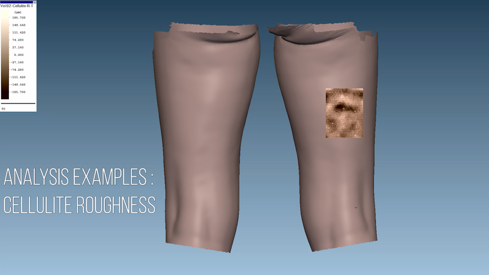
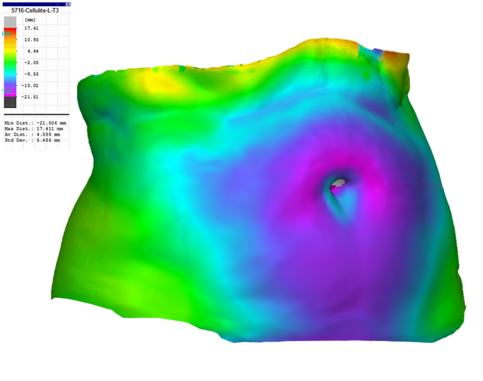
Capturing larger body regions to evaluate volume changes, circumferences, cellulite evaluation and tracking, or slimming effects.
Useful in clinical trials or research labs studying topical or systemic interventions.
Repeatable Clinical Trials
Positioning benches (e.g., VisioHOP) ensure consistent and reproducible scans.
The AEVA-HE² isn’t just another skin camera — it’s a true 3D metrology instrument tailored for high-precision, high-resolution skin and body surface analysis, with rich quantitative outputs that researchers and labs need.
Together with the positioning bench, like the VisioHOP, and the AEVA Software, they create a unified workflow that supports true 3D surface capture, automated analysis, and high reproducibility.
They transform complex skin morphology into reliable, quantitative data suitable for advanced cosmetic research, dermatological studies, and clinical evaluation.
Compared to other systems, AEVA-HE² offers real geometric depth data rather than rendering a 3D look from 2D images — a critical distinction for scientific rigor and repeatable measurement.
AEVA-HE² sets the standard for true 3D skin imaging,
quantitative analysis, and scientific reproducibility

Discover EOTECH’s full range of skin research Instruments at Skinlabs and see how advanced measurement tools can elevate your next research. |
PUBLICATIONS
SKIN RESEARCH & TECHNOLOGY
Shaiek A, Monot M, Rubert V, Cornillon C, Vicic M, Decocq G, Flament F. Applications of the new Aeva-HE™ imaging system: Its link with the visual evaluation of facial wrinkles and its potential in screening tensile products. Skin Res Technol. 2023 Dec;29(12):e13512. doi: 10.1111/srt.13512. PMID: 38081798; PMCID: PMC10713488.
PUBMED
Shaiek A, Monot M, Rubert V, Cornillon C, Vicic M, Flament F, Decocp G, Servant JJ, Koeller G, Lille C. In vitro and in vivo validation of a new three-dimensional fringe projection-based device (AEVA-HE) dedicated to skin surface mapping.
Skin Res Technol. 2023 Feb;29(2):e13209. doi: 10111/srt.13209. PMID: 3674700; PMCID: PMC10155841.
INTERNATIONAL JOURNAL OF PEPTIDE RESEARCH AND THERAPEUTICS
Campiche, R., Jackson, E., Laurent, G. et al. Skin Filling and Firming Activity of a Hyaluronic Acid Inducing Synthetic Tripeptide. Int J Pept Res Ther26, 181-189 (2020). https://doi.org/10.1007/s10989-019-09827-1


Comments