


Lymphedema Management: From Prevention to Early Detection and Monitoring with Quantitative Skin and Tissue Assessment
- Emma Danciu
- May 1
- 9 min read
Lymphedema remains a chronic and often under-diagnosed condition, affecting millions of patients across oncology, surgery, and vascular care. Despite its long-term impact, early-stage lymphedema frequently goes unnoticed, as subtle changes in tissue fluid and structure occur before visible swelling develops.
This diagnostic gap highlights a critical need for objective, quantitative assessment methods that enable clinicians to move beyond subjective evaluation. By integrating precise skin and tissue measurement into clinical practice, healthcare professionals can improve prevention strategies, detect lymphedema earlier, and monitor progression with greater accuracy over time.
Traditionally, lymphedema assessment has relied on visual inspection, limb circumference measurements, and manual palpation. While widely used, these methods are inherently subjective and often lack the sensitivity needed to detect early or subtle changes in tissue composition.
As a result, clinicians may miss the window for early intervention, when treatment is most effective. This limitation underscores the growing importance of objective, reproducible measurement techniques capable of capturing both fluid accumulation and structural tissue changes which are key indicators in the onset and progression of lymphedema.
Understanding What Needs to Be Measured in Lymphedema
Lymphedema is a multifactorial condition that involves more than visible swelling. To accurately assess and monitor its progression, clinicians must consider two fundamental components: tissue fluid accumulation and structural tissue changes. Capturing both dimensions is essential for a comprehensive and clinically meaningful evaluation.
Tissue Fluid Accumulation: The Earliest Indicator
One of the earliest signs of lymphedema is an increase in local tissue water content, reflecting impaired lymphatic drainage. These subtle fluid shifts often occur before any visible or measurable change in limb volume, making them difficult to detect with traditional assessment methods.
Quantifying tissue water provides critical insight into subclinical lymphedema, enabling earlier diagnosis and more proactive intervention.
Structural Tissue Changes: Progression Toward Fibrosis
As lymphedema progresses, persistent fluid accumulation leads to structural alterations within the skin and subcutaneous tissue. This includes the development of fibrosis, characterized by increased tissue stiffness and reduced elasticity.
These changes are associated with more advanced stages of the disease and can negatively impact treatment outcomes if not identified and managed early. Assessing tissue mechanics is therefore essential for understanding disease severity and chronic progression.
What is Lymphedema and How Does It Relate to Skin Fibrosis?
Lymphedema is a chronic condition characterized by the accumulation of lymphatic fluid in the skin and underlying tissues, resulting in swelling, heaviness, and discomfort, most often in the arms or legs.
While visible swelling is the hallmark of lymphedema, the condition often begins sub clinically, with subtle increases in tissue fluid that are difficult to detect without objective measurement.
Over time, persistent fluid accumulation can trigger skin and subcutaneous tissue changes, leading to fibrosis, or the hardening and thickening of the tissue. Fibrosis occurs as a response to chronic lymphatic congestion and inflammation, which stimulates collagen deposition and reduces tissue elasticity.
This combination, fluid overload plus fibrosis, represents the progressive nature of lymphedema, where early-stage swelling evolves into structural tissue changes that are more challenging to manage.
When Do Lymphedema and Skin Fibrosis Occur?
Both lymphedema and skin fibrosis typically appear under circumstances where lymphatic drainage is impaired. Common scenarios include:
Oncologic surgery or radiation therapy: For example, patients undergoing breast, gynecologic, or head and neck cancer treatment may experience lymph node removal or radiation-induced lymphatic damage.
Chronic venous insufficiency or vascular disorders: Poor circulation can lead to lymphatic overload in the lower limbs.
Trauma or infection: Repeated infections, cellulitis, or traumatic injury can impair lymphatic function.
Genetic or congenital conditions: Primary lymphedema can result from lymphatic malformations present at birth or emerging during adolescence.

In the United States, up to 1 in 5 women (20–30%) develop lymphedema after breast cancer surgery, making it a common and often under-diagnosed complication of cancer treatment.
As lymphatic drainage becomes impaired, progressive fluid accumulation and fibrosis can increase arm volume by 20–40% or more, and in severe cases, nearly double the size of the affected limb, significantly impacting mobility and quality of life.
While early detection and intervention can reduce swelling within a few months, lymphedema recovery and stabilization typically take 6 months to 2 years, especially when fibrotic tissue has developed. Because lymphedema is a chronic condition, ongoing monitoring and management are essential to control progression and maintain long-term outcomes.
Who is Affected?
Lymphedema and secondary fibrosis can affect a broad range of patients, but some populations are particularly vulnerable:
Cancer survivors: Breast, gynecologic, and prostate cancer patients are at highest risk of secondary lymphedema.
Post-surgical or post-radiation patients: Any surgery involving lymph node removal or tissue disruption can trigger lymphatic dysfunction.
Individuals with chronic vascular or lymphatic conditions: Those with venous insufficiency, obesity, or infections may develop gradual limb swelling and tissue changes.
Pediatric or congenital cases: Rare genetic conditions can cause primary lymphedema in children and adolescents.
Understanding the interplay between fluid accumulation and fibrosis is crucial for early detection and proactive management. When both factors are monitored together, clinicians can identify subtle changes before visible swelling develops, enabling timely intervention and better long-term outcomes.
Early Signs of Lymphedema and the Limitations of Traditional Assessment
Lymphedema often begins silently, with subtle changes in tissue fluid and skin elasticity that are not immediately visible. Patients may notice heaviness, mild tightness, or slight swelling in an arm or leg, but these early signs are frequently overlooked.
By the time swelling becomes apparent, structural tissue changes, including fibrosis, may already be underway, making management more challenging and less effective.
Why Traditional Methods Fall Short
Conventional lymphedema assessment typically relies on:
Visual inspection: Detects only noticeable swelling; subclinical changes remain hidden.
Limb circumference or volume measurement: Measures gross changes, but small fluid shifts go undetected.
Manual palpation: Subjective and highly dependent on examiner experience.
These methods are often insensitive to early-stage lymphedema, leading to delayed diagnosis and missed opportunities for prevention. For patients at high risk, such as cancer survivors or post-surgical patients, this delay can accelerate the progression toward chronic swelling and tissue fibrosis.
The Need for Objective, Quantitative Assessment
Early detection is only possible when clinicians can measure both tissue fluid accumulation and structural changes with precision and reproducibility. Quantitative skin and tissue assessment enables:
Detection of subclinical lymphedema before visible swelling occurs
Monitoring of fibrotic tissue changes over time
Data-driven decision-making for preventive and therapeutic interventions
Personalized patient management that reduces complications and improves long-term outcomes
By integrating objective measurement into routine care, clinicians can shift from reactive treatment to proactive prevention, stopping lymphedema progression before it becomes irreversible.
A Comprehensive Approach: Measuring Both Tissue Fluid and Stiffness
Effective lymphedema management requires more than just observing swelling. Because the condition involves both fluid accumulation and structural tissue changes, a comprehensive assessment must capture both dimensions.
Tissue Fluid: Detecting Subclinical Lymphedema
Changes in local tissue water content are often the first indicator of lymphatic dysfunction. Subtle fluid shifts occur before visible swelling, signaling that the lymphatic system is under stress. By measuring tissue fluid quantitatively, clinicians can identify subclinical lymphedema, intervene earlier, and reduce the risk of progression to chronic swelling or fibrosis.
Tissue Stiffness: Monitoring Fibrotic Changes
As lymphedema progresses, persistent fluid retention can lead to skin and subcutaneous tissue fibrosis, characterized by increased stiffness and decreased elasticity.
Monitoring these structural changes is essential to:
Evaluate disease progression
Assess severity of chronic lymphedema
Adjust therapeutic strategies for optimal outcomes
Why Both Measurements Matter
Focusing on only one parameter provides an incomplete picture. Measuring fluid without assessing tissue stiffness can miss long-term structural changes, while assessing stiffness alone may detect fibrosis too late for preventive intervention.
By combining fluid and tissue mechanics assessment, clinicians gain a multidimensional view of lymphedema progression, critical for early detection, personalized treatment, and long-term monitoring.
Applying Quantitative Assessment in Clinical Practice
For clinicians seeking a proactive, data-driven approach to lymphedema management, combining measurements of tissue fluid and tissue stiffness offers a complete picture of disease progression.
This dual assessment enables healthcare professionals to detect subtle changes early, track structural tissue alterations over time, and tailor interventions to each patient’s unique needs.
Tissue Fluid Assessment: By precisely measuring local water content in the skin and subcutaneous tissue, clinicians can identify subclinical lymphedema before visible swelling occurs.
Early detection allows for timely preventive strategies, reducing the risk of chronic progression.
Tissue Stiffness Assessment: Evaluating tissue elasticity and firmness provides insight into fibrotic changes that develop as lymphedema progresses.
Monitoring these structural changes helps clinicians understand disease severity, evaluate treatment effectiveness, and adjust therapies for optimal patient outcomes.
Clinical Benefits of a Combined Approach
Implementing both measurements in routine practice supports:
Early Detection and Prevention: Capture changes before swelling becomes noticeable.
Personalized Monitoring: Track both fluid and fibrosis over time for data-driven decisions.
Improved Patient Outcomes: Reduce complications, accelerate recovery, and optimize long-term management.
Evidence-Based Intervention: Provide objective documentation to guide therapy adjustments and validate treatment efficacy.
By integrating these complementary assessments, clinicians can shift from reactive to preventive care, making lymphedema management more precise, proactive, and patient-centered.
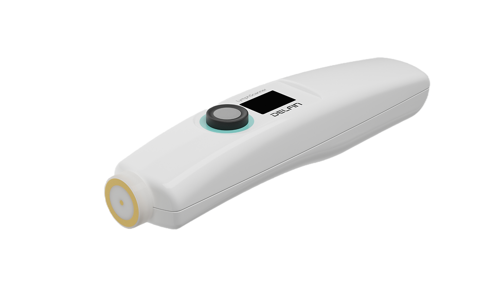
Introducing the LymphScanner™ and SkinFibroMeter™ |
Modern lymphedema management relies on objective, quantitative assessment to detect early changes and track disease progression. Two complementary tools, LymphScanner™ and SkinFibroMeter™, enable clinicians to capture a complete and precise picture of tissue health.
LymphScanner™: Measuring Tissue Fluid Accumulation
LymphScanner™

Measurement
in any position

Measurement
of any part of the body
Technology & Principle
The LymphScanner™ uses Tissue Dielectric Constant (TDC) measurement, a non-invasive method that quantifies local tissue water content.
A small sensor applied to the skin sends a low-level electromagnetic signal (no heat, no feeling), and the tissue’s dielectric properties are measured to determine the Percentage of Water Content (PWC) in both superficial and deeper layers.
Clinical Significance
This allows for subclinical detection of fluid accumulation before visible swelling occurs. It provides objective, reproducible results, overcoming the limitations of traditional methods like circumference measurement or visual inspection.
Unique Advantage
Unlike other devices, the LymphScanner™ captures both free and bound water, offering a more comprehensive assessment of early-stage lymphedema.
BENEFITS OF THE LYMPHSCANNER™ |
Highly sensitive measurement of lymphedema and edema localized in any site | Rapid spot and scanning measurements virtually at all body sites |
Early detection and tracking of lymphedema, with a 2% detection variance accuracy | Force-controlled probe touching skin |
Assess if treatment is working | Totally non-invasive, non-gel measurements, no disposables |
Measures both free and bound water | Fully portable with rechargeable battery |
Percentage Water Content (PWC) displayed | FDA-cleared and CPT coded |
SkinFibroMeter™: Assessing Tissue Stiffness and Fibrosis

SkinFibroMeter™

measures force (N) against indurated subcutis

Measurement
in any position and
on any part of the body
Technology & Principle
The SkinFibroMeter™ employs a precise mechanical indentation system to measure tissue induration.
By applying controlled pressure to the skin and subcutaneous tissue, it quantifies stiffness, enabling detection of fibrotic changes associated with chronic lymphedema.
Clinical Significance
Since subcutaneous fibrosis is situated typically at 2-4 mm under the skin surface, the sensor has to detect the fibrotic or indurated area through the skin.
This objective evaluation of tissue mechanics helps clinicians monitor fibrosis progression, assess disease severity, and guide therapy decisions.
Unique Advantage
Compared to subjective palpation or less sensitive devices, the SkinFibroMeter™ provides highly reproducible, quantitative data, even in early or subtle tissue changes.
BENEFITS OF THE SKINFIBROMETER™ |
Contact pressure-controlled force measurement against fibrous or indurated tissue | Instantaneous measurement (<0.5 seconds) |
Does not alter skin structure | Detects subtle tissue changes early |
Measures force (N) against indurated subcutis (at depth> 2 mm) | Theory of indentation measurement based on material science and 3D modeling of skin and subcutis |
Any position/Anywhere on the body | Fully portable indentation instrument |
Why Using Both Instruments Together Is Crucial
Lymphedema is a multidimensional condition: early fluid accumulation may exist without visible swelling, while tissue fibrosis can develop silently over time. Using either measurement alone provides an incomplete picture.
By combining the LymphScanner™ and SkinFibroMeter™, clinicians gain a full-spectrum, data-driven view of tissue health, enabling:
Early detection of subclinical lymphedema
Monitoring of both fluid and fibrosis simultaneously
Personalized, proactive interventions
Objective tracking of treatment effectiveness over time
This integrated, dual-technology approach is unique in clinical practice, shifting care from reactive management to preventive, precision-driven lymphedema care.
Conclusion: Why Combining Fluid and Fibrosis Assessment is Essential for Optimal Lymphedema Care
We all know lymphedema is a complex, progressive condition, and its impact on patients’ quality of life can be profound. The condition evolves along two critical dimensions: fluid accumulation in the tissue and structural changes such as skin fibrosis.
Both of these symptoms are serious and life-changing, yet they often develop silently in the early stages, making timely detection and intervention crucial.
By combining quantitative tissue fluid measurement with the LymphScanner™ and tissue stiffness assessment with the SkinFibroMeter™, clinicians gain a comprehensive, objective view of a patient’s tissue health.
This dual approach ensures that both subclinical lymphedema and early fibrosis are identified before visible symptoms appear, enabling proactive management and more effective preventive strategies.
The benefits of this integrated assessment extend across the entire patient care continuum:
Prevention: Detect early tissue changes and implement interventions to reduce the risk of disease progression.
Accurate Assessment: Obtain reliable, reproducible measurements of both fluid accumulation and fibrosis for informed clinical decision-making.
Personalized Care: Tailor therapy plans to each patient based on objective data, optimizing outcomes and reducing complications.
Long-Term Monitoring: Track subtle changes over time to evaluate treatment efficacy, adjust interventions, and maintain patient health.
Ultimately, combining these two complementary Instruments empowers clinicians to deliver educated, evidence-based care, shifting lymphedema management from reactive treatment to precision-driven prevention and monitoring.
Patients benefit from earlier detection, better-guided interventions, and improved long-term outcomes, ensuring that both fluid overload and fibrosis are managed effectively and compassionately.
With this dual-measurement strategy, healthcare professionals are equipped to maximize patient health, quality of life, and confidence in care, making it a crucial advancement in modern lymphedema management.
Measure both. Act early. Improve outcomes.
Bring precision & prevention to every patient with lymphedema.
Discover Delfin’s full range of skin research Instruments at Skinlabs and see how advanced measurement tools can elevate your next research. |
PUBLICATIONS |
SWANSEA UNIVERSITY – DEPARTMENT OF BIOMEDICAL ENGINEERING
DITCHFIELD, MARK. Mechanical Assessment of the Skin Through a Multiple Probe Framework to Improve the Assessment and Management of Lymphoedema. Swansea University, Wales, UK, 2024. https://crona.swan.ac.uk/Record/cronfa68319
SAGE JOURNALS
Zaleska MT, Krzesniak NE. Lymphatic Vascular Insufficiency and Focal Edema in Early Stages of Noncancer-related Lymphedema. Lymphatic Research and Biology. 2023;21960 :585-593. doi:10.1089/lrb.2023.0008
SPRINGER NATURE
Yu, Z. (2021). Skin Fibrometer and Lymph Scanner. In: Liu, N. (eds) Peripheral Lymphedema. Springer, Singapore. https://doi.org/10.1007/978-981-16-3484-0_35
SCIENCE DIRECT
Jiajia CHEN, Li WANG, Linghua HAN, Ningfei LIU, Ziyou YU. Therapeutic Efficacy of Complex Decongestive Therapy in the Treatment of Elephantiasis of the Lower Extremities. Chinese Journal of Plastic and Reconstructive Surgery, Volume 2, Issue 1, 2020, Pages 40-62, ISSN 2096-6911, https://doi.org/10.1016/S2096-6911(21)00007-8.


Comments