


Why Multi-Angle Facial Imaging Is Transforming Dermatology and Cosmetic Science
- Emma Danciu
- 1 day ago
- 10 min read
One Facial Photo Is Never Enough
In dermatology and cosmetic science, the smallest skin detail can make the biggest difference.
Skin condition monitoring. Less redness after mesotherapy. A slight improvement in wrinkle depth. A reduction in pore visibility. More even skin tone. Reduced sebum production. These subtle changes are often the difference between a product that performs and one that fails clinical validation.
Yet for years, many skincare brands, dermatologic clinics, CROs, and research laboratories relied on standard photography methods that were highly inconsistent. Variations in lighting, facial positioning, camera angle, shadows, and operator technique could completely alter the appearance of the skin.
The result? Unreliable comparisons, subjective assessments, and difficulty proving treatment efficacy.
Today, the industry is moving toward a more scientific approach: high-precision multi-angle facial imaging.
The Hidden Complexity of Facial Skin Analysis
Human skin is not flat. The face is a highly contoured three-dimensional structure with constantly changing angles, shadows, and textures.
A wrinkle visible from one angle may disappear from another, sebum shine changes depending on light reflection, pigmented spots can appear darker or lighter depending on illumination, pores and skin texture become more or less visible depending on camera positioning.
This is why capturing the face from multiple standardized angles is critical for accurate dermatologic and cosmetic evaluation.
By imaging the face from several perspectives: front, left, right, and oblique views, researchers and clinicians gain a much more complete understanding of the skin’s condition and treatment evolution.
Why Multi-Angle Facial Imaging Matters in Dermatologic and Cosmetic Research
1. Objective Proof of Product Efficacy
Consumers increasingly expect scientific evidence behind skincare claims.
Whether evaluating:
mesotherapy outcomes
acne and blackhead treatments
anti-wrinkle formulations
radiance-enhancing products
sebum-regulating treatments
pore-minimizing solutions
depigmenting products

High-precision imaging provides visual documentation that supports quantitative analysis and clinical conclusions.

Instead of relying solely on subjective grading scales, companies can monitor measurable visual changes over time.
2. Standardization Improves Reproducibility
One of the biggest challenges in skin photography is reproducibility.
Even a few degrees of head rotation or a slight lighting variation can dramatically alter:
wrinkle visibility
shine intensity
pigmentation appearance
pore contrast
skin homogeneity
Standardized multi-angle imaging systems help ensure that every image is captured
under identical conditions:
same positioning
same distance
same lighting
same exposure
same angles
This consistency is essential for longitudinal studies and before/after comparisons.
3. Better Visualization of Skin Topography
Different skin concerns become more visible under different viewing angles.
For example:
crow’s feet are better visualized from oblique views
cheek pores may appear more pronounced under side lighting
facial radiance changes depending on light reflection geometry
pigmentation irregularities may become more visible under controlled illumination
Capturing the face from multiple angles allows researchers to fully assess skin
topography and surface characteristics.
4. Supporting Quantitative Skin Analysis and Dedicated Imaging Software
Modern skin imaging increasingly relies on advanced software capable of extracting
quantitative information from highly standardized photographs.
To generate reliable and comparable datasets, imaging systems must provide:
standardized image acquisition
repeatable positioning
controlled lighting conditions
reproducible facial orientation
high-resolution image capture
Multi-angle facial imaging significantly improves the quality and consistency of
software-based skin analysis for applications such as:
wrinkle assessment
pigmentation and age spot evaluation
skin radiance and homogeneity analysis
pore quantification
blackhead visualization
redness and vascular appearance assessment
skin texture grading
sebum-related evaluations
Capturing the face from multiple perspectives allows subtle skin characteristics to be
documented more completely and consistently, minimizing variability between visit
and improving longitudinal follow-up.
For cosmetic brands, laboratories, CROs, and dermatologic research teams, image
precision directly impacts the reliability of clinical documentation and quantitative
analysis. The more standardized and reproducible the images, the more robust the data
generated for efficacy studies, product claims, and skin evaluation protocols.
How Facial Photography Was Done Before Specialized Imaging Studios
Before dedicated facial imaging systems became available, skin photography was often surprisingly improvised.
Researchers and clinicians commonly used:
standard DSLR cameras
handheld flashes
inconsistent room lighting
manually positioned subjects
operator-dependent framing
This created several major problems.
A participant might slightly tilt their head differently between visits. The lighting intensity could vary from one session to another. Shadows could artificially exaggerate or hide wrinkles. Even camera distance changes could distort facial proportions.
As a result, comparing “before” and “after” images became difficult and sometimes scientifically questionable.
In some cases, image inconsistencies were larger than the treatment effects themselves.
Some Facial Imaging Challenges in Cosmetic Science
Skin Reflects Light Like a Complex Optical Surface | Sebum and skin hydration dramatically influence how light reflects off the skin. Oily skin produces stronger specular reflections, which can affect perceived radiance and texture. |
A Tiny Change in Lighting Can Change Wrinkle Visibility | Wrinkles are highly dependent on shadow formation. Even minimal lighting shifts can make wrinkles appear deeper or smoother. |
Human Eyes Are Surprisingly Subjective | Two trained evaluators may interpret the same skin improvement differently. High-precision imaging helps reduce subjective bias. |
Same Face Can Look Different From Different Angles | Some pigmentation spots, pores, and vascular structures become visible only under certain viewing perspectives or illumination geometries. |
Consistent Imaging Is Essential for Clinical Credibility | Regulatory documentation and scientific publications increasingly require standardized imaging protocols to support product claims. |
Why High-Precision Facial Imaging Helps Develop Better Products
Accurate imaging is not only about marketing claims. It directly contributes to innovation in dermatology and cosmetic science.
When researchers can precisely visualize subtle skin changes, they can:
better understand treatment mechanisms
optimize formulations
identify responder profiles
refine application protocols
improve treatment personalization
accelerate R&D decision-making
Ultimately, better imaging leads to better products, more reliable studies, and improved outcomes for patients and consumers.
From Theory to Practice:Enter the CBright Facial Imaging System |
Understanding the importance of standardized multi-angle facial imaging is one thing. Implementing it reliably in real clinical and cosmetic environments is another challenge entirely.
This is precisely where the CBright system, along with its dedicated Revelare software, becomes a major asset for dermatologic and cosmetic research.
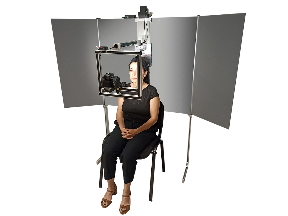
Rather than functioning as a simple camera setup, CBright was specifically designed as a dedicated facial imaging studio engineered for reproducible, high-precision skin photography.
For cosmetic brands, CROs, dermatology clinics, and research laboratories, the system helps transform facial photography from a subjective visual exercise into a standardized scientific process.


What Is CBright?
The CBright is a compact facial imaging system developed specifically for dermatologic follow-up, cosmetic efficacy studies, aesthetic medicine, clinical research, and quantitative skin analysis.
Its purpose is to acquire highly reproducible facial photographs under fully controlled conditions.
Unlike conventional photography systems, CBright combines:
standardized positioning
controlled LED illumination
reproducible camera geometry
multi-angle acquisition
guided facial alignment
optional polarized imaging

The result is a highly consistent imaging workflow capable of documenting subtle skin changes with exceptional reproducibility.
This is essential when studying:
Acne and vitiligo monitoring
mesotherapy outcomes
redness and vascular appearance
anti-wrinkle treatments
radiance enhancement
age spots and pigmentation
skin homogeneity
pores and blackheads
sebum-regulating products
Why Standardization Is So Important
In dermatologic and cosmetic imaging, inconsistency is the enemy of reliable data.
Even tiny variations in:
head position
chin elevation
facial rotation
camera distance
illumination angle
can significantly alter the appearance of:
wrinkles
skin texture
shine
pigmentation
pores
skin relief
A wrinkle may suddenly appear deeper simply because shadows changed. Skin radiance may seem improved due to different light reflections rather than actual treatment efficacy.
CBright addresses these issues by creating a controlled imaging environment where every parameter is standardized.
The system incorporates precise positioning assistance, angular presets, facial centering guidance, stable illumination conditions, and reproducible acquisition protocols.
This dramatically improves before-and-after comparability throughout long-term studies.
Why Multi-Angle Facial Imaging Changes Everything
One frontal photograph alone cannot fully represent the complexity of facial skin.
The skin features behave differently depending on the viewing angle and lighting geometry.
CBright allows acquisition from multiple standardized facial angles (0°, 30°, 45°, 60° and 90°), helping researchers visualize the skin more comprehensively.
Multi-angle facial imaging improves both visual clinical assessment and quantitative software analysis.
For dermatologic and cosmetic companies, this means more reliable efficacy documentation and stronger scientific validation.
The Technology Behind CBright
One of CBright’s greatest strengths is the integration of several technologies specifically optimized for skin imaging reproducibility.
Controlled LED Illumination
Lighting consistency is critical in skin analysis.
CBright integrates homogeneous LED illumination designed to minimize variability between imaging sessions.
Controlled lighting is essential for evaluating radiance, skin homogeneity, pigmentation, redness, sebum shine, and skin texture.
Without standardized lighting, visual differences may reflect environmental variations rather than actual biological changes.
Dedicated Positioning System
CBright includes positioning assistance systems that help maintain reproducible facial alignment throughout longitudinal studies.
This reduces operator-related variability and improves acquisition consistency over time.
The system helps standardize:
. head orientation
. facial centering
. acquisition angles
. camera-subject geometry
This level of repeatability is extremely valuable for clinical studies requiring rigorous image comparison.
Polarized Imaging Capabilities (Including Cross-Polarization)
CBright also supports polarized imaging modes, including both parallel polarization and cross-polarization, which are essential tools in modern dermatologic and cosmetic skin analysis.
Polarized light is used to control how light interacts with the skin surface, allowing clinicians and researchers to distinguish between surface reflections and subsurface skin information. This is critical when evaluating subtle treatment effects that may otherwise be masked by glare or lighting variability.
a) Parallel Polarization
In parallel polarization, the emitted and captured light share the same orientation. This configuration preserves more surface reflections and is particularly useful for analyzing:
. skin gloss and radiance
. sebum distribution and oily zones
. superficial skin texture
. surface shine variations
This mode is especially relevant in cosmetic studies focused on skin glow, hydration appearance, and oil control treatments.
b) Cross-Polarization
In cross-polarization, the emitted and captured light are oriented perpendicularly. This effectively reduces surface glare and suppresses specular reflections, allowing deeper skin structures to become more visible.
This mode is widely used for evaluating:
. pigmentation and age spots
. redness and vascular appearance
. inflammatory skin conditions
. subsurface color variations
. texture irregularities beneath the surface
Cross-polarization is considered a cornerstone in dermatologic imaging because it reveals the true chromatic properties of the skin, independent of surface shine.


Why Polarization Is a Game-Changer in Skin Analysis
By combining both polarization modes, CBright enables a dual-layer analysis of the skin:
Parallel polarization → surface behavior (shine, sebum, radiance)
Cross-polarization → subsurface conditions (pigmentation, redness, vascularity)
This dual capability is particularly important for cosmetic and dermatologic studies where improvements are often subtle and multi-dimensional.
For example, an anti-aging treatment may reduce wrinkle depth (structural change), while also improving radiance (optical surface change), and reducing redness (vascular change).
Each of these effects is best evaluated using different imaging modalities, which CBright integrates within a single standardized system.
What Does Revelare Do?
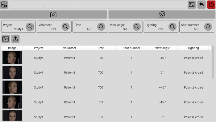
Revelare, the integrated software, manages the entire imaging workflow from acquisition to study organization.
It can organize studies, projects, users, volunteers or patients, acquisition sessions, and imaging protocols.
It guides operators step-by-step through standardized acquisition procedures, helping reduce human error and variability.
This is particularly important in clinical studies where reproducibility is critical.
One of the largest sources of variability in skin imaging is not necessarily the camera itself, but inconsistencies in image acquisition procedures. This is where Revelare improves standardization and scientific reliability.
Revelare addresses this problem directly and helps standardize:
image capture sequences
acquisition angles
session organization
imaging parameters
data traceability
Each image is automatically linked to:
patient or volunteer information
study session
acquisition conditions
imaging metadata

Revelare improves study consistency, traceability, reproducibility, and operational efficiency.
For CROs and cosmetic laboratories managing large datasets, this represents a considerable advantage.
Why Raw Image Integrity Matters
In cosmetic and dermatologic research, image authenticity is essential.
Post-processed images can unintentionally smooth wrinkles, alter pigmentation, modify contrast or exaggerate treatment outcomes.
Revelare helps preserve image integrity by maintaining standardized acquisition workflows and reliable image management practices.
This contributes to stronger scientific credibility and more trustworthy efficacy documentation.
Why CBright Is a Major Asset for Dermatologic and Cosmetic Industries
For cosmetic brands and dermatologic researchers, CBright and Revelare offer far more than photography.
They provide:
scientific rigor
reproducible documentation
standardized imaging protocols
improved clinical credibility
optimized workflow management
stronger product validation
CBright and Revelare are a valuable tool for clinical research, as it allows researchers to create panels of multiple volunteers captured at identical view angles. This standardization greatly facilitates image comparison, streamlines workflow, and improves study efficiency.
As skincare claims become increasingly evidence-based, standardized imaging systems are becoming indispensable tools for modern cosmetic science.
Because in today’s dermatologic and cosmetic industries, proving efficacy requires more than beautiful photographs.
It requires measurable, reproducible, scientifically controlled imaging data.
CBright
Compact Photographic Studio for Reproducible & Standardized Face Photos
CBright
Precision Lighting for multi-angle imaging with optional cross-polarization
Parallel-polarized imaging to quantify brightness & surface properties
Image acquisition fully automated by the Revelare software
Multiple angles (0°, 30°, 45°, 60° and 90°)
Low-intensity LED illumination enables subjects to keep their eyes open


Capture the face at multiple angles and change the future of its skin.
Discover EOTECH’s full range of skin research Instruments at Skinlabs and see how advanced measurement tools help you do more for science. |
SCIENTIFIC CONTEXT |


Comments